Revolutionizing healthcare with accurate EVD placement
Improving safety, speed, and accuracy
THE SOUNDPASS SOLUTION
A new approach to EVD placement
Our team is building the world’s first ultrasound and reconstruction algorithm capable of 3D imaging from an astounding 1.65mm diameter transducer. This will give neurosurgeons the tools they need for successful EVD placement in even the most challenging situations.
Why SoundPass?
The Urgency
Due to the life-threatening nature of rapid brain swelling after traumatic brain injury (TBI), neurosurgeons will place EVDs to remove Cerebral Spinal Fluid (CSF) to prevent brain herniation, permanent disability, and death, which may happen in a matter of hours if not done promptly.
Seeing Blind
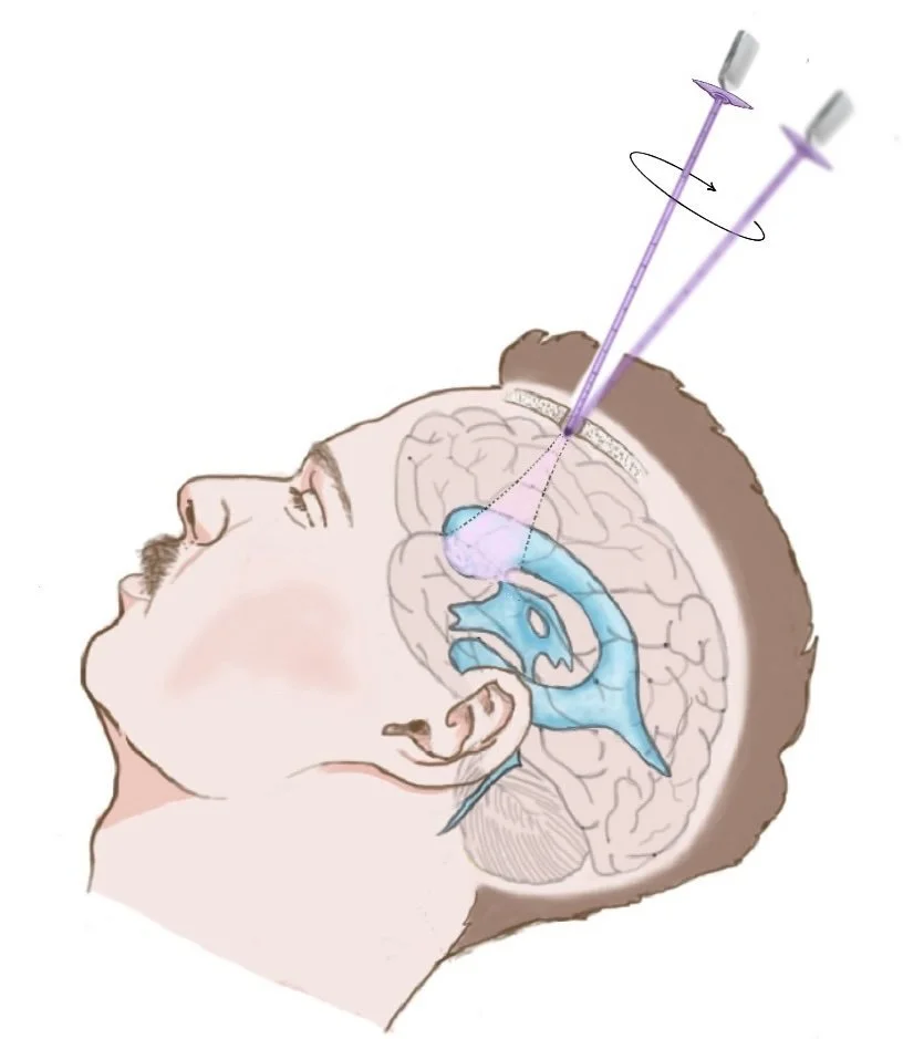
With this urgency, placement of EVDs are often done at the bedside (outside of the operating room) based on external anatomy alone (“blind approach”). Neurosurgeons have no way to see inside the skull to place EVDs in their target location (Right Lateral Ventricle).
The Cost
Surgeons often require 2–8 (or more) attempts through healthy brain tissue to find the ventricle if squished – if at all. The current standard of care (“blind placement”) for EVDs is associated with high error, morbidity, and unnecessary mortality, which we believe shouldn’t be the case.

THE SOUNDPASS MISSION
“To save human life and the quality thereof by enhancing the accuracy, speed, precision, and availability of life-saving external ventricular drain (EVD) placement.”
Want to get involved?
3 Pillars of Success
Ultrasound Tipped Stylet
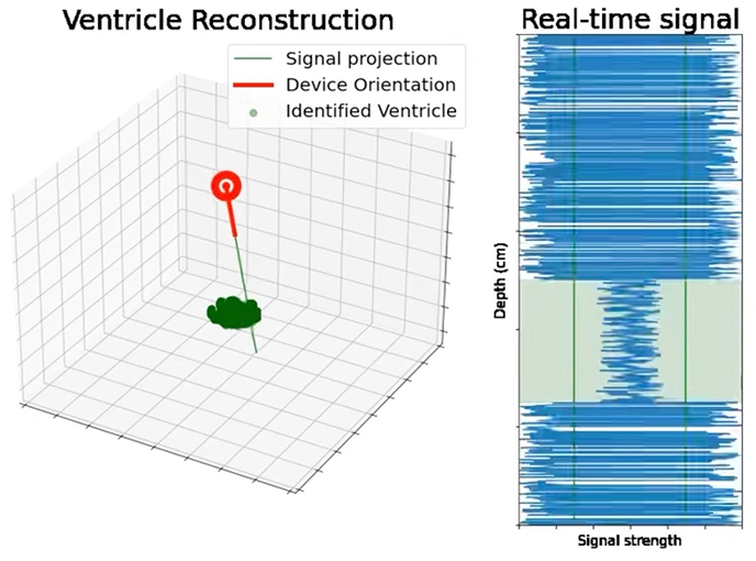
Our stylet replaces the current steel stylet used to give rigidity to the external ventricular drain. A novel transducer with a 1.65mm diameter custom-designed to measure at the depth of the ventricles provides the raw data needed for a useful reconstruction.

Echolucent Catheter
Our proprietary echo-lucent catheter not only allows for imaging to the depth of the ventricle, but also enhances and focuses the ultrasound beam produced by the stylet.

3 Dimensional Reconstruction
Our 3D reconstruction algorithm identifies and reconstructs the ventricles, allows the neurosurgeon to choose their trajectory, and shows them their trajectory, so they can follow the placement all the way into the ventricle.


